IL-33
Interleukin-1 (IL-1) family, such as IL-1α/β and IL-18, has important functions in host defense, immune regulation, and inflammation. IL-33, a member of the IL-1 family, that shows to induce T helper (Th) type 2 responses by signaling through the IL-1 receptor-related protein ST2 (IL-1R4), an orphan member of the IL-1 receptor family. Similarly to IL-1α/β and IL-18, IL-33 is synthesized as a 31 kDa precursor protein has been shown to be cleaved by caspase-1 in vitro. In vivo, IL-33 induces the expression of IL-4, IL-5, and IL-13 and leads to severe pathological changes in mucosal organs. IL-33 has been originally identified as NF-HEV, which is a nuclear factor preferentially expressed in high endothelial venules. IL-33 may function as both a proinflammatory cytokine and an intracellular nuclear factor involved in transcriptional regulation.
IL-33 is also a member of the IL-1 family; however it differs from IL-1β in its activation control mechanism. Like IL-1α, IL-33 is present in the nucleus under normal conditions. Although the function of IL-33 as a nuclear factor remains elusive, its binding to the nucleosomal surface to suppress transcription has been demonstrated by in vitro experiments.
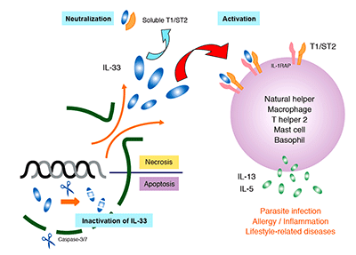
IL-33 is expressed in a variety of cells (including endothelial, epithelial, and fat cells) and tissues (such as the stomach, lungs, skin, lymph nodes, and kidneys). Since IL-33 is released during cell necrosis or tissue damage, is also suggested that IL-33 alerts the immune system after disruption to endothelial or epithelial cells has occurred. Its up-regulated expression is reported in the mouse brain and spinal cord. IL-33 is has been implicated in diseases ranging from parasitic infection and allergic diseases to arthritis, diabetes, inflammatory bowel disease, SLE, and Alzheimer’s disease.
Specific IL-33 Involvement:
Necrosis:
Full-length IL-33 is released extracellularly and activates the immune cells expressing IL-33 receptors. At the inflammation site, IL-33 undergoes limited proteolysis by the proteases released from neutrophils and other cells, and limited proteolysis results in its enhanced activity.
Apoptosis:
IL-33 is cleaved by activated caspase-3 and caspase-7, which terminates its inflammation-inducing ability.
Infections:
Full-length IL-33 released extraceullarly acts on diverse leucocytes to induce production of primarily Th2-type cytokines and plays a role mainly in defense mechanisms against parasite infection. A team from Keio University discovered the presence in the mouse visceral fat tissues of natural helper (NH) cells that secrete a large amount of Th2-type cytokines through IL-33 action. This has become a hot topic because the part of the system that provides protection from parasite infection has been elucidated.
Research Grade IL-33 Antibodies Available
Code No. | Species Reactivities | Applications | Clonality | Notes |
---|---|---|---|---|
M161-3 | Mouse | WB, IP | Monoclonal | Research Grade |
M138-3 | Human | WB, IP | Monoclonal | Research Grade |
PM033 | Human | WB, IHC | Polyclonal | Research Grade |
Functional Grade IL-33 Antibodies Available
Code No. | Species Reactivities | Applications | Clonality | Notes |
---|---|---|---|---|
M187-3 | Mouse | WB, NT | Monoclonal | Functional Grade |
M188-3 | Mouse | WB, NT | Monoclonal | Functional Grade |
IL-33 ELISA kit Available
Code No. | Species Reactivities | Applications |
---|---|---|
7650 | Human | ELISA |
Citations
Neutralization
Ohno T et al. Paracrine IL-33 stimulation enhances lipopolysaccharide-mediated macrophage activation. PLoS One 6, eOhno, Tatsukuni, et al. “Paracrine IL-33 Stimulation Enhances Lipopolysaccharide-Mediated Macrophage Activation.” PLoS ONE, vol. 6, no. 4, 2011, doi:10.1371/journal.pone.0018404.
Bunting, Melissa M., et al. “Interleukin-33 Drives Activation of Alveolar Macrophages and Airway Inflammation in a Mouse Model of Acute Exacerbation of Chronic Asthma.” BioMed Research International, vol. 2013, 2013, pp. 1–10., doi:10.1155/2013/250938.
Shadie, A M, et al. “Ambient Particulate Matter Induces an Exacerbation of Airway Inflammation in Experimental Asthma: Role of Interleukin-33.” Clinical and Experimental Immunology, vol. 177, no. 2, 2014, pp. 491–499., doi:10.1111/cei.12348.
Function
Saigusa, Ryosuke, et al. “Fli1-Haploinsufficient Dermal Fibroblasts Promote Skin-Localized Transdifferentiation of th2-like Regulatory T Cells.” Arthritis Research & Therapy, vol. 20, no. 1, 2018, doi:10.1186/s13075-018-1521-3.
Additional Publications
Angulo, Evelyn L., et al. “Comparison of IL-33 and IL-5 Family Mediated Activation of Human Eosinophils.” PLOS ONE, vol. 14, no. 9, 2019, doi:10.1371/journal.pone.0217807.
Chacon, Nathalie, et al. “Implications of Helminth Immunomodulation on Covid-19 Co-Infections.” Life Research, vol. 4, no. 3, 2021, p. 27., doi:10.53388/life2021-0502-309.
Li, Xiaochen, et al. “Interleukin-33, a Potential Cytokine Expressed in the Tumor Microenvironment Is Involved in Antitumor Immunotherapy through Facilitating CD8+ T Cells.” Journal of Interferon & Cytokine Research, vol. 38, no. 11, 2018, pp. 491–499., doi:10.1089/jir.2018.0069.
Murakami, Shokei, et al. “An Epidermal Keratinocyte Homogenate Induced Type 2 and Proinflammatory Cytokine Expression in Cultured Dermal Cells.” Journal of Dermatological Science, 2022, doi:10.1016/j.jdermsci.2022.04.002.
Okragly, Angela J, et al. “Generation and Characterization of Torudokimab (LY3375880): A Monoclonal Antibody That Neutralizes Interleukin-33.” Journal of Inflammation Research, Volume 14, 2021, pp. 3823–3835., doi:10.2147/jir.s320287.