
- Bright orange fluorescence
- High pH stability
- Monomer/dimer
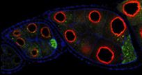
CoralHue® Kusabira-Orange (KO1), from the stony coral Fungia concinna (Kusabira-ishi in Japanese), absorbs light maximally at 548 nm and emits orange light at 561 nm. KO1 rapidly matures to form a fluorescent dimeric complex. KO1 can be used to mark cells or to report gene expression without problems stemming from protein aggregation.
CoralHue® monomeric Kusabira Orange (mKO1) maintains the brilliance and pH stability of the parent protein. mKO1 can be used to label proteins or subcellular structures or for FRET analysis.
CoralHue® mKO2 is a mutant of mKO1 that features rapid maturation. mKO2 can be used to label proteins or subcellular structures or for reporter assays.
Performance and Use
Kusabira-Orange are useful for labeling organelle.
Kusabira-Orange is easily expressed and detected in a wide range of organisms. It has been demonstrated in nucleoplasm, plasma membrane, endoplasmic reticulum and mitochondrial targeting signal models.
Kusabira-Orange is a useful protein for expression into various species.
 |
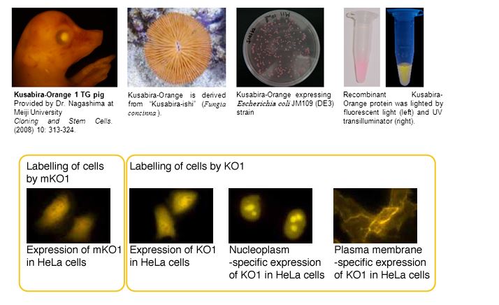
K01 TG pig – Provided by Dr. Nagashima, Meiji University, Cloning and Stem Cells. (2008) 10: 313-324. |
 |
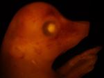
mKO1 fused with Vasa protein expressed in Drosophila ovary (red). – Provided by Dr. Nakamura RIKEN |
 |
The olfactory bulb of zebrafish – Provided by Dr. Miyasaka and Dr. Yoshihara, Laboratory for Neurology of Synapse, BSI, RIKEN |
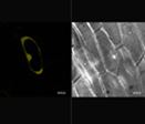 |
KO1 expressed in the epidermis of onion – Provided by Dr. Iida and Dr. Hoshino, the National Institute of Basic Biology (NIBB). |
Citations
- Karasawa S, Araki T, Yamamoto-Hino M, Miyawaki A, A green-emitting fluorescent protein from Galaxeidae coral and its monomeric version for use in fluorescent labeling; J Biol Chem. 278, 34167-34171 (2003) PMID: 12819206
- Ebisawa T, Yamamura A, Kameda Y, Hayakawa K, Nagata K, Tanokura M. Crystallization and preliminary X-ray analysis of a monomeric mutant of Azami-Green (mAG), an Aequorea victoria green fluorescent protein-like green-emitting fluorescent protein from the stony coral Galaxea fascicularis. Acta Crystallogr Sect F Struct Biol Cryst Commun. 65(Pt 12):1292-5. (2009) PMID: 20054132
Fluorescent Properties
- Kusabira-Orange1 excitation (8K)
- Kusabira-Orange1 emission (8K)
- mKusabira-Orange1 excitation (8K)
- mKusabira-Orange1 emission (8K)
- mKusabira-Orange2 excitation (4K)
- mKusabira-Orange2 emission (8K)
Note: The file is in a tab-delimited text format. It contains values of the wavelength (0.5nm spacing) and brightness (fluorescence intensity peak value normalized to 1). Use a spreadsheet program to create a spectrum that will help you in choosing the appropriate excitation filter, dichroic mirror and fluorescence filter.
| Characteristic | K01 | mK01 | mK02 |
| Oligomerization | Dimer | Monomer | Monomer |
| Number of Amino Acid | 218 | 218 | 218 |
| Excit./Emiss. maxima (nm) | 548/561 | 548/559 | 551/565 |
| Molar Extinction Coefficient (M-¹cm-¹) | 73,700 (548 nm) | 51,600 (548 nm) | 63,800 (551 nm) |
| Fluorescence quantum yield | 0.45 | 0.60 | 0.62 |
| Brightness*¹ | 33.2 | 31.0 | 39.6 |
| pH sensitivity | pKa<5.0 | pKa<5.0 | pKa=5.5 |
| Cytotoxicity*² | Not observed | Not observed | Not observed |
| Resistance to PFA fixation | Not tested | Not tested | +++ |
*1Brightness: Molar Extinction Coefficient ×Fluorescence Quantum Yield / 1000
*2Toxicity when expressed in HeLa cells
Recommended Antibodies
CoralHue® KO1, mKO1 and mKO2 can be recognized using antibodies as shown below.
- Anti-monomeric Kusabira-Orange 1 for WB, Code No. M104-3M
- Anti-monomeric Kusabira-Orange 2 for WB, IP, IC and IH, Code No. M168-3M
- Anti-monomeric Kusabira-Orange 2 for WB, IP, IC and IH, Code No. PM051M
WB: Western blotting, IP: Immunoprecipitation, IC: Immunocytochemistry, IH: Immunohistochemistry