
Useful for Single Cell Analysis of Cell and Organelle Movement
- Photoconvertible fluorescent protein
- Tracking of movement of single cells and organelles
- High Sensitivity and Excitation to UV light
- Monitor Morphology of Neurons
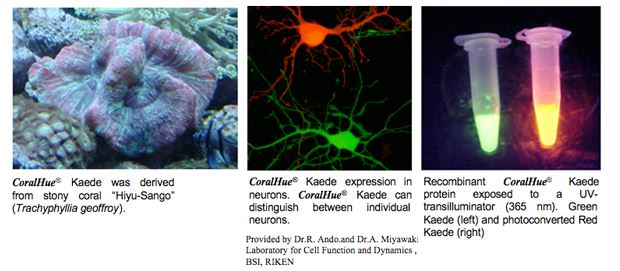
The fluorescent protein Kaede gene was isolated from the stony coral Trachyphylia geoffroyi (Hiyu-sango in Japanese).
Kaede means maple leaf in Japanese. CoralHue® Kaede protein emits bright green fluorescence that can irreversibly convert to red. The red fluorescence is comparable in intensity to the green and is stable under usual aerobic conditions. The green-to-red conversion is highly sensitive to irradiation with UV or violet light (350-410 nm). Maximal illumination results in a 2,000-fold increase in the ratio of red-to-green signaling. The excitation lights used to elicit red or green fluorescence do not induce the photoconversion.
This property provides a simple and powerful technique for regional optical marking. Cell (Ref.2,3), organelle (Ref.4) or protein dynamics (Ref.5,6) have been reported.
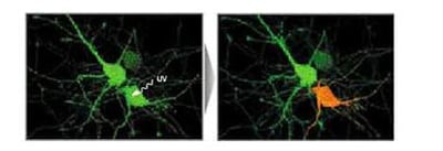
Irradiation with UV to Kaede expressing cells converts the green fluorescence to red, which leads to the diffusion of red colored Kaede throughout the Kaede expressing cell. The photoconversion can be induced gradually by intermittent irradiation.
Expression of CoralHue® Kaede in neurons. Without irradation with UV, Kaede protein emits green fluorescence which visualize the morphology of neurons (Left). Irradation with UV to the cell body of one neuron elicits the green-to-red fluorescence conversion specifically in the irradiated cell, by which neighboring neurons can be distingueshed by color (Right).
(Images are provided by Dr. Miyawaki Atsushi,
Laboratory for Cell Function and Dynamics, BSI, RIKEN)
Citations
- Ando R, Hama H, Yamamoto-Hino M, Mizuno H, Miyawaki A. An optical marker based on the UV-induced green-to-red photoconversion of a fluorescent protein. Proc Natl Acad Sci U S A. (2002) 99(20):12651-6. PMID: 12271129
- Tomura M, Yoshida N, Tanaka J, Karasawa S, Miwa Y, Miyawaki A, Kanagawa O. Monitoring cellular movement in vivo with photoconvertible fluorescence protein “Kaede” transgenic mice. Proc Natl Acad Sci U S A. (2008) 105(31):10871-6. PMID: 18663225
- Hatta K, Tsujii H, Omura T. Cell tracking using a photoconvertible fluorescent protein. Nat Protoc. (2006) 1(2):960-7. PMID: 17406330
- Watanabe W, Shimada T, Matsunaga S, Kurihara D, Fukui K, Shin-Ichi Arimura S, Tsutsumi N, Isobe K, Itoh K. Single-organelle tracking by two-photon conversion. Opt Express. (2007) 15(5):2490-8. PMID: 19532486
- Schmidt A, Wiesner B, Weisshart K, Schulz K, Furkert J, Lamprecht B, Rosenthal W, Schulein R. Use of Kaede fusions to visualize recycling of G protein-coupled receptors. Traffic. (2009) 10(1):2-15. PMID: 18939954
- Leung KM, Holt CE. Live visualization of protein synthesis in axonal growth cones by microinjection of photoconvertible Kaede into Xenopus embryos. Nat Protoc. (2008) 3(8):1318-27. PMID: 18714300
Fluorescent Properties
Kaede Spectrum Data Files
- Kaede-green excitation (8K)
- Kaede-green emission (8K)
- Kaede-red excitation (16K)
- Kaede-red emission (8K)
Note: The file is in a tab-delimited text format. It contains values of the wavelength (0.5nm spacing) and brightness (fluorescence intensity peak value normalized to 1). Use a spreadsheet program to create a spectrum that will help you in choosing the appropriate excitation filter, dichroic mirror and fluorescence filter.
Filter Set for Kaede
Filter set for Kaede is available from Opto Science, Inc.
For more information, please see Opto Science, Inc. webpage
*1Brightness: Molar Extinction Coefficient ×Fluorescence Quantum Yield / 1000
*2Toxicity when expressed in HeLa cells
Transgenic Animals
Kaede transgenic mouse
The Kaede transgenic mouse was developed by Dr. Miwa (Ref.2). Kaede is a photoconvertible fluorescence protein that changes from green to red upon exposure to violet light. The photoconversion of intracellular Kaede has no effect on cellular function. Thus, Cell dynamics can be monitored with the photoconverted Kaede protein.
Provided by Dr.Y. Miwa
Department of Molecular Pharmacology, Graduate School of Comprehensive Human Sciences, University of Tsukuba
Visualization of cell movements and the tracing of neuronal shapes using Kaede transgenic zebrafish (Ref.3)
Recommended antibodies
CoralHue®Kaede can be recognized using antibodies as shown below.
- Anti-Kaede for WB, Code No. M125-3M
- Anti-Kaede for IP, Code No. M106-3M
- Anti-Kaede for WB, Code No. PM012M
WB: Western blotting, IP: Immunoprecipitation