
- Bright green fluorescence
- High pH stability for minimal loss of signal
- Monomer/tetramer
- Useful for labeling proteins or subcellular structures
CoralHue® Azami-Green (AG), cloned from the stony coral Galaxea fascicularis (Azami-sango in Japanese), absorbs light maximally at 492 nm and emits green light at 505 nm. AG is stable in the standard biological pH range and does not show a significant loss of fluorescent signal, unlike other fluorescent proteins such as EGFP. And AG also matures rapidly to form tetramers that are brighter than EGFP.
CoralHue® monomeric Azami-Green (mAG1) maintains the brightness and pH stability of the parent protein AG. mAG1 can be used for labeling proteins or subcellular structures.
Performance and Use
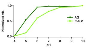
AG is resistant to acidity
AG shows high resistance in a wide range of biological pH. This allows AG to be used to identify cells or to report gene expression without being influenced by pH change. The pH dependence of both CoralHue® AG and mAG1 was identical to that of its absorption spectra.
AG and mAG1 are useful for labeling organelle.
AG and mAG1 performance in fusions has been demonstrated in the nucleoplasm, plasma membrane, ER and mitochondria-targeting signal models. We offer these constructs as targeting vector series.
mAG1 is available for protein labeling
mAG1 performance in fusion has been demonstrated in p80 coilin, SC35 and fibrillarin.azami7
Fluorescent Properties
Azami Green (AG) spectrum data files (text files)
- AG excitation (8K)
- AG emission (8K)
- mAG1 excitation (8K)
- mAG1 emission (8K)
Note: The file is in a tab-delimited text format. It contains values of the wavelength (0.5nm spacing) and brightness (fluorescence intensity peak value normalized to 1). Use a spreadsheet program to create a spectrum that will help you in choosing the appropriate excitation filter, dichroic mirror and fluorescence filter.
*1Brightness: Molar Extinction Coefficient x Fluorescence Quantum Yield/1000
*2Toxicity when expressed in HeLa cells
Recommended antibodies
CoralHue® AG and mAG1 can be recognized using antibodies as shown below.
- Anti-monomeric Azami-Green 1 for WB, Code No. M102-3M
- Anti-Azami-Green for WB, Code No. PM011M
- Anti-monomeric Azami-Green 1 for WB, IP, IC and IH. Code No. PM052M
WB: Western blotting, IP: Immunoprecipitation, IC: Immunocytochemistry, IH: Immunohictochemistry
Publications
- Karasawa S, Araki T, Yamamoto-Hino M, Miyawaki A, A green-emitting fluorescent protein from Galaxeidae coral and its monomeric version for use in fluorescent labeling; J Biol Chem. 278, 34167-34171 (2003) PMID: 12819206
- Ebisawa T, Yamamura A, Kameda Y, Hayakawa K, Nagata K, Tanokura M. Crystallization and preliminary X-ray analysis of a monomeric mutant of Azami-Green (mAG), an Aequorea victoria green fluorescent protein-like green-emitting fluorescent protein from the stony coral Galaxea fascicularis. Acta Crystallogr Sect F Struct Biol Cryst Commun. 65(Pt 12):1292-5. (2009) PMID: 20054132